Electrodes, leads & wires
A practical guide to ecg monitoring and recording.
A striking example of what can happen if you change the limb electrode positions.
Several A&E nurses have told me of carefully placed chest electrodes being removed by ccu staff, and replaced with chest electrodes in the wrong positions. This may be because ccu are using the EASI™ 12 lead monitoring system - see below.
An excellent paper "Resting 12-Lead ECG Electrode Placement and Associated Problems" has been brought to my attention. Available from the Society for Cardiological Science and Technology web site. It contains many example ecg's to help you spot incorrect electrode placement.
Settling arguments
The most frequent question I get asked, in or out of the classroom, is "Where should the electrodes be placed". But before I answer, let's consider some basic principles, and see how the answers depend on a number of factors.
OK, if you don't want to read it all, you can go straight to the most common question "where to put the limb electrodes for a 12 lead ecg".
Always remember that when someone insists on doing things a certain way, they can usually find that someone somewhere has written a paper on it. I've had to suffer working with anaesthetists who insist that a particular electrode must go in a particular position. At the time I knew it did not matter, but I always preferred a quiet life in theatre, so I just let them get on with it. ;-)
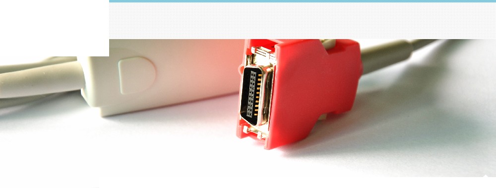
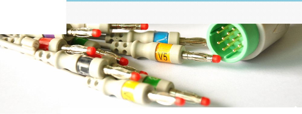
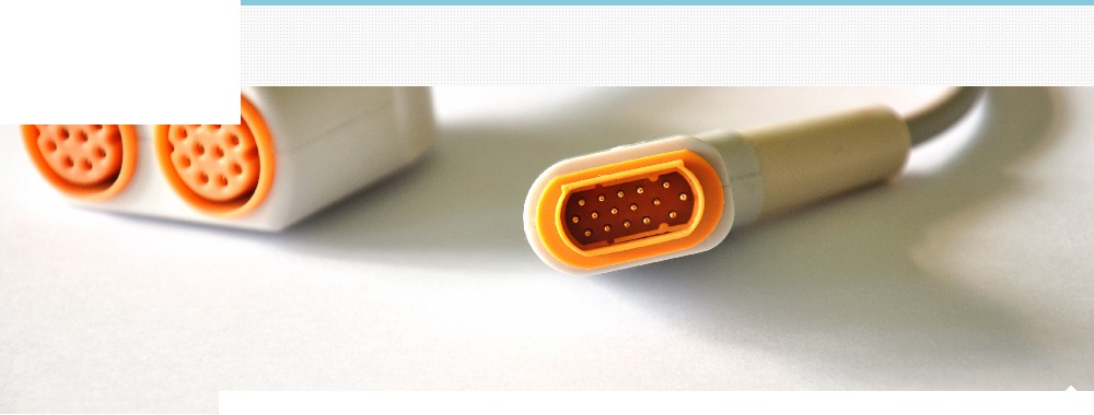
Leads and wires - why 6+4=12
Between the patient and the ecg machine is a patient cable, and this is divided into a number of different coloured wires, maybe 3, 4 or 5 for monitoring purposes, or 10 wires for a 12 lead ecg.
A lead is a view of the electrical activity of the heart from a particular angle across the body, obtained by using different combinations of these wires. Leads are pictures, you cannot point to one of the wires or electrodes on the patient and say "this is lead so and so", to say this you would have to point to a "group" of wires.
To obtain a 12 lead ecg you would have 4 wires attached to each of the limbs, and six wires placed around the chest, 10 wires in total but you get 12 "leads" or pictures. In a similar way a combination lock may have 50 numbers but it has thousands of possible combinations.
Leads and wires (part 2) - how 5 wires can give you 12 leads
Philips Medical Systems produce the EASI™ 12 lead system.
This uses 5 electrodes and "derives" the 12 leads from these. The electrode positions are:
- Upper sternum (just below sternal angle)
- Lower sternum (fifth intercostal space)
- Right midaxillary line (fifth intercostal space)
- Left midaxillary line (fifth intercostal space)
- Ground (can be anywhere)
But let's now leave such specialised systems and go back to basics.
How the standard 12 leads "look" at the heart
To measure any electrical activity you need at least two electrodes (a positive and a negative) in order to form an electrical circuit. If using defibrillator paddles to obtain a trace, you are using this principle. So we have one electrode "looking" between itself and the other electrode. By changing the position of either of these electrodes we alter the angle at which we are viewing any activity.
Important point, no matter which lead is being used to "look" at the heart, the electrode doing the looking is always a single electrode. But the "other" electrode could be a single electrode or a number of other electrodes joined together (electrically) to form a reference point.
The six chest leads are simple enough, each of the six chest electrodes looks from that point on the chest wall, the six wires give you six different views. The "other" electrode in each of these views, is created by the machine "joining together" the limb connections, thus making the centre of the body the reference point.
The six limb leads work differently, leads I, II and III are each making use of a pair of electrodes, with one electrode looking between itself and the other as shown below. Leads aVR, aVL and aVF each make use of all the limb connections to the patient, but again one single electrode is doing the looking (relative to the others).
When you try to think of how the limb leads look at the heart imagine the views as taken from positions around a clock face (as facing the patient).
With electrodes on the right arm (red), left arm (yellow) and left leg (green)
- Lead I uses red and yellow, yellow is looking from 3 o'clock.
- Lead II uses red and green, green is looking from 5 o'clock.
- Lead III uses yellow and green, green is looking from 7 o'clock.
- Lead aVR is looking from the red wire, across to between yellow and green, 10 o'clock.
- Lead aVL is looking from the yellow wire, across to between red and green, 2 o'clock.
- Lead aVF is looking from the green wire, across to between yellow and red, 6 o'clock.
When considering all the above leads, and the wires that are involved. Notice that you must have the limb electrodes connected to the patient in order to record the six chest leads. But you do not need the chest electrodes connected in order to record the limb leads.
My thanks to Dr Bruce Graham of Charles Sturt University NSW Australia for contributing suggestions concerning the limb and chest leads.
Different coloured wires
It's important that you know if you're using a European or American patient cable for monitoring because the colours of the wires will differ as shown below.
All references on this page are based on the european (IEC) cable colours.
| Monitoring cable connections |
| Europe
Red
Yellow
Green
Black
White |
Connect to:
Right Arm
Left Arm
Left Leg
Right Leg
Chest |
U.S.A.
White
Black
Red
Green
Brown |
| Individual chest leads |
| White / Red
White / Yellow
White / Green
White / Brown
White / Black
White / Violet |
C1 / V1
C2 / V2
C3 / V3
C4 / V4
C5 / V5
C6 / V6
|
Brown / Red
Brown / Yellow
Brown / Green
Brown / Blue
Brown / Orange
Brown / Purple
|
Different numbers of wires
A 3 wire cable (red, yellow, green) or (red, yellow, black) can only give you a choice of limb leads.
A 4 wire cable (red, yellow, green, black) can only give you a choice of limb leads.
A 5 wire cable (red, yellow, green, black, white) will give you limb leads plus a chest lead (using the white wire - usually placed in the V1 position). This is a versatile monitoring cable, if you have one and lend it out - you may not get it back.
A 10 wire cable is for recording a 12 lead ecg.
What type of ecg are you doing?
Do you have the patient on a monitor, or are you recording a 12 lead ecg? Your needs will differ.
Monitoring
If monitoring for changes in rhythm, you are interested in what the ventricles and atria are doing. As long as you can see clearly big and small waves of activity from these two areas, you can work out the rhythm. Choose the lead that gives the best picture of what you are interested in; the size/shape of the p wave and/or the qrs. it's often lead II, lead I or a chest lead, but your choice may be limited by injuries, dressings or the patients position. But where should the electrodes be placed, wrist, forearm, upper arm, shoulder, lower or upper leg or even on the torso in a square box arrangement - it does not matter, getting a good signal is more important than precise electrode positioning when monitoring rhythm. It's common to place the red wire on the right shoulder, yellow on the left shoulder and green (or black) at the apex of the heart, it may not look much like a triangle but it will produce a fairly good lead I and II when you turn the lead selector switch. Then we come to the old argument about "bony" or "muscular" area beneath the electrode. Well it's six of one to half a dozen of the other; bone is not such a good conductor but is less likely to produce muscular interference near to the electrode, muscle is a better conductor but more likely to introduce interference. Remember that if you turn up the gain (increase the size of the trace) on the monitor, you also increase the size of any interference.
Common sense would tell you that the further you are from the heart the smaller the waves of activity will be, but there will be no difference in time intervals; p-r interval etc. So get in close, but not so close that you might interfere with attempts at defibrillation or cardioversion. And a patient monitored in bed with electrodes on their wrists and ankles would not be as happy as one with electrodes on their torso.
If monitoring for ischaemic changes, you will be interested in a particular area of the heart. Usually a chest lead is used (you can get closer to the area involved), try to get the correct position (more on this later), but more importantly be consistent with the position if the electrodes are changed.
So why do people put electrodes in different places for monitoring?
I mentioned using different leads to obtain different views, in other words turning the lead selector switch on the monitor to a different lead position. Most monitors give you leads I, II and III. BUT some simple monitors have no lead selector switch, they're usually fixed to give you lead I (but check the operators manual). These machines often have a cable with 3 wires; red, yellow and black. I mentioned above that for simple monitoring the red is on the right shoulder, yellow on the left shoulder and black at the apex. Now if the machine is giving you a lead I trace, it's using red and yellow with yellow doing the "looking" or detecting, the black is acting as an earth connection. If you put the yellow down at the apex, and move the black up to the left shoulder, you've changed the angle across the chest, in fact you've fooled the machine into giving you a lead II trace. The position of the black wire is not that important in this situation, that's why in the operating theatre you will often see the black wire attached to an electrode on the patients forehead or arm, to keep it out of the way.
Instead of turning a switch to obtain different leads, we are changing the position of the electrodes. But don't do this if your monitor has a lead selector switch or you will screw up the views that leads I II and III give you.
Important point
I once had a prolonged argument with a senior cardiographer who insisted that the black wire never records anything and is only an earth connection. This is true with 12 lead ecg's, but is not always so with monitoring.
If your monitor has 3 wires; red, yellow, black, and has a lead selector switch, and the shape of the complex changes when you turn the switch to different positions.
You are using red and yellow for lead I, red and black for lead II, yellow and black for lead III.
Other monitoring leads
We saw above that moving electrodes to different positions can "fool" the monitor into providing a different lead or view.
A popular monitoring lead is MCL1 (stands for modified central lead one), this fools the monitor into giving you a simulated lead V1, here's how. With a 3 wire cable; put the red wire on the left shoulder just below the clavicle, black (or green) on the right shoulder, yellow in 4th intercostal space on the right sternal border (the V1 position), if there's a lead selector switch set it to lead I. Alternatively, if you put the black (or green) in the V6 electrode position, turning the lead selector switch to lead II would give you a simulated lead V6.
MCL1 is a useful monitoring lead, it usually gives good waves for rhythm analysis and is the best lead for identification of bundle branch blocks.
Many different configurations are possible using a 3 wire system, and some of these have been forgotten such as; CS5 (MCR5), CM5, CB5, CC5. To make sense of any of these modified leads you must consider what lead has been selected on the monitor, and which wires it therefore uses, in particular which wire is doing the "looking".
Telemetry and Holter monitoring can offer single, dual or multiple lead configurations which make use of combinations of limb leads I, II and III with modified chest leads.
If you make a paper recording of a rhythm strip you must write on it what lead (or simulated lead) was used.
Electrodes
Please please please do not leave electrodes attached to the patient cable, or put neatly arranged rows of electrodes "ready to go" on the arrest trolley unless they are used on a daily basis. The electrode gel will dry out and be useless. I once attended an "arrest" on a ward where the resus trolley was used perhaps once every couple of months, every electrode had to be thrown away because they had dried out. If you do find yourself in this situation, break out some normal saline and put a spot on the dried out gel pad, it should work. You may like to know that Lewes published a paper in the British Heart Journal in 1965 showing that ketchup, mayonnaise, toothpaste and K-Y jelly all produced equally good results as electrode gel.
The twelve lead ecg
Let's split this into 6 chest leads and 6 limb leads.
The six chest leads
The electrodes for the chest leads MUST go in the standard positions:
- V1 - Fourth intercostal space, right sternal border.
- V2 - Fourth intercostal space, left sternal border.
- V3 - Midway between V2 and V4.
- V4 - Fifth intercostal space, left midclavicular line.
- V5 - Level with V4, left anterior axillary line.
- V6 - Level with V4, left mid axillary line.
In female patients electrodes are NEVER placed on top of the breast unless you cannot gain access to the normal position. If you do have to move onto the breast, write it on the recording.
However, having said that, I'm grateful to PWharmby@aol.com for telling me about the following:
"Breast tissue appears to have a practically negligible effect on ECG amplitudes, and in women, the placement of chest electrodes on the breast rather than under the breast is recommended in order to facilitate the precision of electrode placement at the correct horizontal level and at the correct lateral positions."
Rautaharju PM, Park L, Rautaharju FS, Crow R. A standardized procedure for locating and documenting ECG chest electrode positions: consideration of the effect of breast tissue on ECG amplitudes in women. J Electrocardiol. 1998 Jan;31(1):17-29.
V1 is the important starting point, if you get this wrong the whole lot will be wrong. It can be difficult locating the fourth intercostal space. The best way is to run your fingers down the sternum, starting at the heads of the clavicles, until you meet a bony horizontal ridge (the sternal angle or angle of Louis), this is easier to find in male patients. With your finger on this ridge, slide it to the patients right, your finger will drop into an intercostal space, this is the second intercostal space, now move down to the third and then the fourth, here's where you place V1.
The most important aspect of 12 lead ecg recording is consistency of recording technique, but positioning of electrodes comes a close second.
When using the 12 lead ecg to look for evidence of ischaemia or infarction, we are not so much interested in a single 12 lead recording (though it may be conclusive on its own), but how it changes over time - we are comparing recordings. If these records were not all made the same way the reliability of comparison is lost.
If you think the chest electrodes are in the wrong place, and a recording was previously made (say 10 minutes ago). DON'T change the electrodes to the "correct" positions until you have made a final recording using those same electrodes. Then do a "proper" recording and write on it that the electrodes were repositioned.
An extreme example
Say I put an electrode on my nose and one on my big toe, I would record some activity. Then 20 minutes later I make another recording using my nose and big toe. I could compare it to the first, I could say if there were changes. It would be of use.
Once you've placed the chest electrodes and made a recording leave them alone.
Extra chest leads
It's often useful to look further round the heart than V1 to V6. You can do "right chest leads", designated as V1R to V6R - a mirror image of the normal chest leads. Remember that your normal V2 is in fact V1R, and normal V1 is V2R, so to include a "right" set of chest leads just record V3R to V6R.
Posterior leads can be useful. V7 is level with V6 in the left posterior axillary line, V8 is level with V7 at the left midscapular line (often taken as midway between V7 and V9), and V9 is level with V7 at the left spinal border.
If a chest lead is recorded from the tip of the ensiform (xiphoid) process, it is designated with a lower case "e", as in Ve.
Remember that to record these extra leads you would have to reposition some of the wires to the new electrode positions, the machine will not know you have done this. It will still print "V1" or "V6" or whatever next to the tracing, you must cross this out and write the new lead position.
The 15 lead ecg
Some 12 lead ecg machines have the capability of recording some or all of these extra leads. On some models of the Hewlett Packard Pagewriter look at the large plastic block that holds the wires that go off to the limb and chest connections. You may see some extra slots for wires to be plugged in, labelled as C3R etc. Just add the wires and reconfigure the machine setup to print a 15 lead ecg (standard 12 lead plus three extra chest leads).
The six limb leads of the 12 lead ecg
Last, but certainly by no means least. The topic that generates the most arguments, is it wrists arms or shoulders for the arm connections, and ankles thighs or hips for the leg connections..
First an historical perspective. Very early ecg recordings were made by having the patient lie down with the ends of their arms and legs dipped in buckets of salt water (only limb leads were used originally) in order to make the required electrical connections. Then someone came up with the idea of using a saline soaked pad placed around the wrist or ankle, with a metal plate on top - secured by a rubber strap, which can have a wire attached that goes to the recording device - no more buckets. Then we get the idea to replace the saline soaked pad with some conductive jelly between the metal plate and the skin - everyone stays dry, but we still need the rubber strap to hold them in place.
At this point it's worth noting that although the arms and legs can be likened to "wires" hanging off the torso; the impulse that reaches say the right arm has to travel from the right shoulder to get there. It would have required a very "Heath Robinson" Victorian type contraption to strap electrodes to the shoulders and groin, it was just easier to put the connections on the wrists and ankles.
Then we get adhesive electrodes, and everyone goes crazy. It's now realised that you're not limited to wrists and ankles when choosing a site for the electrode. Move closer to the heart (shoulders and groin) and you get a stronger signal, plus you are less likely to pick up skeletal muscular interference or tremor. And female patients don't have to remove their tights.
How this can affect the 12 lead ecg
Analysis of the 12 lead ecg is all about measurements, the height and depth of waves can be important in the diagnosis of certain conditions such as infarction or hypertrophy. Technically there will be a slight difference in size of waves seen in the limb leads if you record a limb lead from a torso connection compared to a limb connection. But this will not affect the complexes in the chest leads.
The following changes in the 12 lead ecg have been shown to occur when the 4 limb electrodes are placed on the torso;
- A shift in the cardiac axis towards the right.
- R wave becomes smaller in lead I.
- Less prominent Q waves in inferior leads.
Bartosik J, Pahlm O, Edenbrandt L, et al. Reconstruction of the standard 12-lead ECG from recordings using nonstandard activity-compatible proximal limb lead positions. J Electrocardiol 1995 Jan; 28(1):33-8.
An example of different limb electrode positions
I'm grateful to Kerry Smith, Clinical Coordinator of the Arrhythmia Technologies Institute for sending me these striking examples of how evidence of an inferior MI can "disappear" by moving the limb electrodes to the torso.
So what are the correct positions for the limb electrodes
According to the American Heart Association, "The electrodes may be placed on any part of the arms or of the left leg as long as they are below the shoulders in the former and below the inguinal fold anterioly and the gluteal fold posterioly in the latter. Any other placement necessary by deformed or missing extremities must be noted on the record."
Pipberger HV, Arzbaecher RC, Berson AS, et al. Recommendations for standardization of leads and of specifications for instruments in electrocardiography and vectorcardiography: Report of the Committee on Electrocardiography, American Heart Association. Circulation 1975;52:11Ð31.
You can not compare an ecg recorded with torso limb lead placement, with a standard ecg.
But you could compare a series of ecg's recorded in such a way.
For the analysis of serial changes (such as in ischaemia / infarction), consistency of recording technique is important.
There may be times when you need to digress from standard technique. Say the patient had a peripheral muscular tremor, you may get a better quality recording by using torso limb lead placement.
If your department/unit/ward/hospital or clinic places the arm connections on the upper arms or shoulders, just make sure everyone does it the same way. Then all recordings are comparable (but not in different parts of the country or the world).
There is a recognised international standard for placement of the limb electrodes (as well as the chest connections), wrists and ankles. If you want to do it different, fine, so long as everyone you work with knows and agrees (that's the hard part).
If you record a 12 lead ecg with any aspect of recording technique differing from the standard (see below), write what you have done on the recording. For example "Torso placement of limb electrodes".
Many practitioners now give patients a copy of their last recorded ecg. This is an excellent idea, that recording will be useful to any future paramedic, nurse, or doctor. But it would be best to record it the standard way; patient supine, arms by their side, chest electrodes in their correct positions, limb electrodes on wrists and ankles, recorded at 25mm/sec, calibrated at 10mm/mV.
Having said all of this, there is bound to be someone somewhere who will disagree with something said.